|
The helical ventricular myocardial
band (HVMB) of Torrent-Guasp, bringing a new light on
perennial problem of global, macroscopic,
three-dimensional functional architecture of the
ventricular myocardium, emerged in early 1970s.
Not perchance, the earliest
recognitions came from outside of Spain.
Professor Sir Donald N. Ross, the
close friend and the great admirer of Torrent-Guasp’s
work, invited him to Guy's Hospital, London, to expose
the HVMB concept, which Jane Somerville subsequently
compared to Leonardo Da Vinci’s masterpiece.
Some years later, in Geneva,
Torrent-Guasp was awarded the Miguel Servetus Prize,
signed by Nobelist, Sir Ernst B. Chain.
Since that time, the original HVMB
concept has considerably evolved, not only as a result
of dedicated work of its creator and his associates, but
also because of fascinating advances in experimental and
imaging technology, supported by powerful computing
hardware and algorithms.
HVMB
Anatomy
Here you may find two review papers
describing current knowledge of HVMB anatomy:
Kocica MJ, Corno AF, Carreras-Costa F, Ballester-Rodes
M, Moghbel MC, Cueva CNC, Lackovic V, Kanjuh VI,
Torrent-Guasp F.
The helical ventricular myocardial band: Global,
three dimensional functional architecture of the
ventricular myocardium.
Eur J Cardiothorac Surg 2006;29(S1):S21-S40.
EJCTS Suppl: Rethinking the
cardiac helix: a structure-function journey.
TOP25 articles: European Journal
of Cardio-Thoracic Surgery (April - June 2006)
Journal impact factor: 1.616 (2004)

Torrent-Guasp F, Kocica MJ, Corno AF, Komeda M,
Carreras-Costa F, Flotats A, Cosin-Aguillar J, Wen H.
Towards new understanding of the heart structure and
function.
Eur J Cardiothorac Surg 2005;27(2):191-201.
TOP25 articles: European Journal
of Cardio-Thoracic Surgery (January - December 2005)
Journal impact factor: 1.616 (2004)
HVMB Dissection
Technique
Preparations
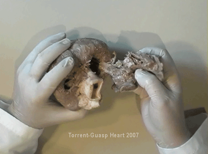
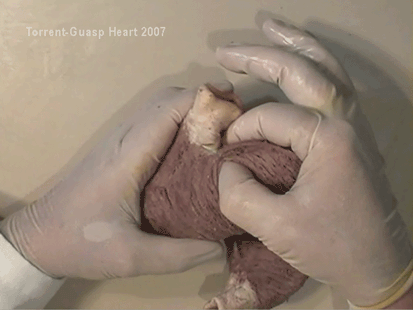
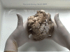
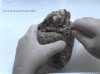
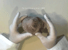
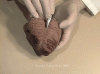
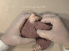
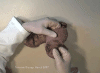
Figures 1-20 are depicting preparation phases
for Torrent-Guasp's anatomical dissection of the HVMB.
The hearts are prepared by simple boiling in water
(without any additive), in order to loosen the
connective tissue. The period of boiling was judged
empirically, on the appearance of fibers and depended
on the size of the specimen - about 10 minutes or less
for a hen heart and up to 2 hours for an adult bovine
heart. After boiling and subsequent cooling (several
hours in refrigerator at 4C), the atria could be
easily removed from the heart. Pulmonary artery and
aorta are trimmed, leaving proximal 2-3cm. The fat
from the atrio-ventricular grooves was removed and all
visible, superficial coronary vessels excised.
Dissection of the myocardial mass is undertaken with
non-toothed forceps, scalpel and scissors. Blunt
dissection by fingers is generally the most
satisfactory way of identifying the direction of the
linear (fiber) and laminar (layer) pathways. Gentle
longitudinal traction was enough to separate long
strips of myocardium, whereas forcible lateral
traction tended to tear the muscle fiber.
Dissection technique
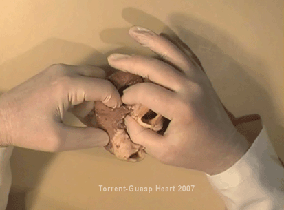
Figures 21-53 are depicting successive steps of
Torrent-Guasp's dissection technique applied for
unraveling the ventricular mass into the HVMB.
After the separation of the pulmonary artery and the
aorta, some superficial fibers (i.e. aberrant fibers)
bridging the anterior interventricular sulcus are
incised in order to move aside the right ventricular
(RV) free wall. By doing so, we arrive to the
posterior linear border of the RV cavity, which is
represented by the linear bottom of the dihedral angle
constituted by the RV free wall and the
interventricular septum. The posterior linear border
of the RV cavity has special importance, since it
points out the only possible trajectory, which would
allow further dissection of the HVMB. The beginning of
this trajectory is exposed by pushing laterally RV
free wall. Following the predominant fiber direction,
we can easily see that this path encircles the LV, up
to the root of the aorta.
By cutting their anchorage with the left fibrous
trigone, we have finished the dissection of the HVMB
basal loop. At this point, it is important to notice
that some fibers (i.e. belonging to the descendent
segment) are sinking into the LV, making the central
fold of the HVMB. Trajectory of these fibers, while
coming down towards the LV posterior wall, is pointing
out an important cleavage plan at level of the
interventricular septum. Namely, at the septal level,
these fibers are crossing the ascendant segment fibers
in a 90-degree angle. At this point, we are able to
see this septal crossing from the LV side.
To continue with dissection, we should come back, to
the site of the previous posterior linear border of
the RV cavity. By pure inspection from the RV side, we
can clearly distinguish two muscular strata. The
deeper belongs to previously described descendent
segment and the more superficial belongs to the
ascendant segment. A right-angle crossing of these
fibers, as described before, is now also visible from
the RV side. The cleavage plan between these two
strata, is the same one we described above, entering
it from the LV side. The top of the line (i.e.
previous posterior linear border of the RV), defined
by these two strata, ends on the aortic root at the
point of it’s attachment to the right fibrous trigone.
To separate described strata, going in between the
vertical (more superficial, ascendant segment) and the
horizontal (deeper, descendent segment) fibers, the
first thing that we should do is to cut-off their
anchorage to the right fibrous trigone. Now we are
able to proceed with the most delicate part of the
dissection, denominated as “dismounting of the aorta”.
Prior to any further description of the dissection
method, it is important to emphasize one fact. The
only firm aortic attachments to the LV are the fibrous
trigones, upon which the aorta leans over the LV
outflow tract. Apart from that, the aortic annulus,
belonging to the right coronary cusp, provides the
additional, weak anchorage of the aorta to the septal
portion of the LV. Thus, by cutting-off these firm and
weak attachments, it becomes possible to dismount the
aorta from the LV. By doing so, we are able to join
two parts of the septal cleavage plan. In this manner,
progressing along the predominant fiber path, we are
able to detach the aorta with fibers belonging to the
ascendant segment from the rest of the LV mass.
Following the same cleavage plan along the predominant
helical fiber path, we are entering the LV cavity,
with fingertips appearing behind the anterior
papillary muscle, at the level of previously mentioned
central fold of the HVMB. If we proceed until we
become able to close the fist, our fingertips would
appear between anterior and posterior papillary
muscle, the former being completely encircled by the
hand.
Finally, we came to the most exciting part of the
dissection, when the HVMB is ready to be
stretched-out. Simple 90-degree rotation around the
apex unravels the apical loop segments. Additional
180-degree rotation around the central fold unravels
the basal and the apical loops of the HVMB. The HVMB
of Torrent-Guasp now appears in its full extent and
beauty, with pulmonary artery at one and the aorta at
the opposite side.
The elegance and astounding simplicity of this
dissection is reflected in the capacity to easily
reverse these unraveling steps, with ready
re-establishment of the well-known three-dimensional
ventricular architecture that existed prior to
beginning of dissection.
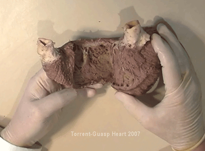
Segmental anatomy of the HVMB
Figure 54 emphasizes four
crucial dissection phases and segmental anatomy
of Torrent-Guasp's HVMB.
The HVMB is divided in two loops, each of them
comprising of two segments. The central 180-degree
fold of the HVMB defines two loops: the
basal loop (from the root of the pulmonary artery
to the beginning of the central fold - i.e. to the
anterior papillary muscle) and the apical loop
(from the beginning of the central fold to the root of
the aorta). Each of these two loops could be further
divided in two segments.
The posterior interventricular sulcus, which coincides
topographically with the posterior linear border of
the RV cavity, divides the basal loop into two
segments: the right
segment - coinciding with the RV free wall;
and the left segment
- coinciding with the LV free wall. It is interesting
to notice here, that the right segment also defines
the outer (non-septal) border of the tricuspid orifice
and the left segment defines the outer (non-septal)
border of the mitral orifice. These borders are common
targets in AV surgical annuloplastic procedures.
The apical loop could be also divided in two
segments. After the 180-degree twist (at the
central fold of the HVMB), fibers
of the
descendant segment,
make a 90-degree turn around the apex,
continuing the fibers of
the ascendant
segment.
Posterior papillary muscle (belonging to the
descendant segment), demarcates the border between the
descendent and the ascendant segments of the HVMB
apical loop.
 |
 |
 |
 |
 |
|
Figure 1 |
Figure 2 |
Figure 3 |
Figure 4 |
Figure 5 |
 |
 |
 |
 |
 |
|
Figure 6 |
Figure 7 |
Figure 8 |
Figure 9 |
Figure 10 |
 |
 |
 |
 |
 |
|
Figure 11 |
Figure 12 |
Figure 13 |
Figure 14 |
Figure 15 |
 |
 |
 |
 |
 |
|
Figure 16 |
Figure 17 |
Figure 18 |
Figure 19 |
Figure
20 |
 |
 |
 |
 |
 |
|
Figure
21 |
Figure
22 |
Figure
23 |
Figure
24 |
Figure
25 |
 |
 |
 |
 |
 |
|
Figure
26 |
Figure
27 |
Figure
28 |
Figure
29 |
Figure
30 |
 |
 |
 |
 |
 |
|
Figure
31 |
Figure
32 |
Figure
33 |
Figure
34 |
Figure
35 |
 |
 |
 |
 |
 |
|
Figure
36 |
Figure
37 |
Figure
38 |
Figure
39 |
Figure
40 |
 |
 |
 |
 |
 |
|
Figure
41 |
Figure
42 |
Figure
43 |
Figure
44 |
Figure
45 |
 |
 |
 |
 |
 |
|
Figure
46 |
Figure
47 |
Figure
48 |
Figure
49 |
Figure
50 |
| |
 |
 |
 |
|
| |
Figure
51 |
Figure
52 |
Figure
53 |
|
| |
|
|
|
|
 |
|
Figure
54 |
|