|
The knowledge is the circle,
surrounded by ignorance. The bigger the circle - the
wider the frontier towards the unknown.
This wisdom helped me a lot, not to be afraid of
ignorance. I was happy once being able to ask questions.
One of these questions bothered me since 1997, and was
related to reasons of better ventricular performance
after mitral valve replacement with preserved
annulo-papillary continuity. Since the earliest
observations by Walton Lillehei, a lot has been written
and said about this phenomenon, but underlying mechanism
remained controversial.
It took me four years to think about it, although, I was
almost sure that the answer is hidden inside the natural
masterpiece of ventricular myocardial architecture.
Unfortunately, I did not keep a record of papers I've
read during these four years, but all in vain. Each time
I thought the answer is close - I realized there was a
missing link, which I could not find in classical
literature.
My ideas about the functional importance of
annulo-papillary continuity and possible anatomical
explanations were not accepted by my colleagues,
although, they did not provide me any alternative
explanation. This kind of situation (I believe known to
many people) gave me additional strength to keep on
searching.
Until the October 2001, my concept was based on
comparisons with elbow biomechanics.

Elbow biomechanics and mitral complex.
In brief - if we cut-off the ulnar insertion of the
biceps brachii muscle, we would not be able to hold
any weight in our hand, in spite of the fact that this
muscle can contract, whenever it is excited.
Therefore, this muscle can perform intrinsic work
- but without preserved insertions on the opposite
arms of the elbow lever - it can not do any
extrinsic work. For the persons with this problem
- this means a great loss of arm function – almost
invalidity.
I've always kept in my mind the figure from the
textbook of anatomy I have used as the medical
student. The figure was by Jane Sands Robb from 1942.
At this moment, I tried to compare the effects of
chordal resection with the resection of biceps’ ulnar
insertion. The only thing that was clear to me, was
the fact - that chordal resection leaves not only
papillary muscle unsupported - but also produces a
similar effect to certain adjacent (and remote) parts
of the ventricular myocardium. I could not know
exactly the extent of this effect.
My erroneous conception was that closed loop - formed
by the papillary muscles, myocardium, mitral annulus,
mitral leaflets and chordae tendineae – has the same
functional explanation as the loop (or triangle)
formed by biceps brachii, radius and ulna. The
concepts of the fibrous skeleton of the heart and the
architecture of the ventricular fibers - had different
meaning for me.
I was desperate, because (being a cardiac surgeon) I
could not organize any kind of anatomical research at
our Medical School. I had an idea to build up a wire
frame with 1x1cm windows and then to start peeling off
myocardial fibers (starting form the epicardium) -
making a photographs from time to time - in order to
produce 3D reconstruction of fiber architecture. My
friends Ivan and Dejan, who are "PC magicians",
encouraged me with information that they will be able to
render such 3D object. I knew that this kind of job is
almost impossible and I started to lose my enthusiasm.
Miracles do not happen in
contradiction to nature, but only in contradiction to
what is known to us of nature.
Saint Augustin
And then came a miracle. I had very old PC (486, 100MHz,
1Gb) and funny Internet connection of 9.4 Kbps. I spent
a nights trying to find something interesting and
related to my "problem" on the Web. You can imagine my
reaction (Figure 2), when I saw the link for the JTCVS
paper:
-
Torrent-Guasp F, Ballester M, Buckberg GD, Carreras F,
Flotats A, Carrio I, Ferreira A, Samuels LE, Narula J.
Spatial orientation of the ventricular muscle band:
physiologic contribution and surgical implications.
J Thorac Cardiovasc Surg. 2001 Aug;122(2):389-92.
The Gospel of Matthew (7:7) - "Ask, and it shall be
given you; seek, and you shall find..."
Of course, I did not wasted my time, and I sent a mail
to corresponding author (i.e. Dr. Manel Ballester-Rodés)
exposing him my problems and asking for his opinion. I
did not have an idea about Francisco Torrent-Guasp,
although he was signed as the firs author of this paper.
Soon after this, came the first reply from Francisco
Torrent-Guasp (Paco). That was the beginning of the most
beautiful period in my career. I felt like "Alice in
Wonderland", confused and delighted at the same time. I
have realized the meaning of freedom and the magnitude
of nature, which were put together in perfect harmony in
every single aspect of Paco's life. It was apparent
simplicity in everything he did - not the primary but
the ultimate simplicity - being the language by which
the nature reveals its secret to very few people.
Here follows the initial
correspondence between Paco and me.
No 1 - Paco:
Dear Dr. Kocica,
Dr. Ballester Rodés has sent to me your message, with
the doubts and questions related with heart structure
and mechanics. I must tell you that I have been
working on these themes during half a century and now,
after those 50 years, I have been able to describe the
ventricular macroscopic structure (by means of the
anatomical definition of the ventricular myocardial
band) and the ventricular mechanics (explaining the
way the ventricles decrease and increase their volume
by means of respective muscular contractions).
Therefore it seems (that is what what general opinion
says) that I have been able to solve the old problem,
stated first time by Erasistratus 2300 years ago, to
show which is the mechanical trick used by the
ventricles to increase their volume by means of a
contraction, apparent incoherent fact since any
contraction usually implicates decrease of dimensions,
retractions, shortening, etc.
Recently, last month of September, has appeared an
article of mine in the REVISTA ESPAÑOLA DE CARDIOLOGIA
(Rev Esp Cardiol 2001 ; 54 : 1091-1102). This article
has been summarized in the JTCVS article that you have
read, in which appears as coauthors Dr. Ballester and
some others. I will send you the translation, into my
very bad English, of this article.
If you want to go further you could come to Dénia and
I will give you any kind of details at the same time
that you will learn to dissect the ventricular
myocardium to evidence the ventricular myocardial
band; I did show it in many places of Europe, USA,
Japan, etc., and soon I am going to do it in Mexico,
Brazil, Uruguay and Argentina. Next March-April the
NIH of USA has organized a Workshop on my work that
will take place in Washington attending about 30 basic
heart research authors.
And now let me try to answer to your questions, as Dr.
Ballester asked me to do.
FIRST QUESTION :
It seems your are worried about the connections of the
ventricular myocardial band with the fibrous skeleton.
First I must tell you that such concept, fibrous
skeleton, was a very unfortunate idea inserted by the
classic anatomists in the mind of people. Only the
aortic and pulmonary artery rings and the two trigons,
are consistent anatomical entities.
The mitral and tricuspid rings are just a line along
which the atria are stuck to the ventricles. It is
more ; most of the ventricular muscular fibers are not
inserted in these two A-V rings, as the classic
anatomists say. The ventricular myocardial band is
inserted, in one end, in the pulmonary artery ring
and, in the other end, in the aortic ring; nothing
more. Not even the trigones or used as a fixed point
by the ventricular fibers. You write in you letter,
that "there is not efficient contraction if muscle is
not attached (supported) somewhere". That is true on
respect the skeletal muscles, as the biceps, but it
does not applies to the circular muscles. The circular
muscles rest, when they contract, on the contents of
the tubular cavity they delimitate, i.e., the circular
muscles of the vessels rest on the blood, the circular
muscles of the bowels rest on the food that has been
eaten, the circular muscles of the bronchia on the
air, the circular muscles of the sphincters on what is
defined by the short tube they define, etc. Therefore
the fulcrum of the circular muscles is a mobile point
(the blood, the food, the air, etc., are running), not
a skeletal fixed point as the one represented by the
insertions of any skeletal muscle in a bone (as the
biceps, triceps, and so on). The circular muscles do
not need any fixed point to perform their activity.
And it happens that the ventricular muscle band
describe two spirals (loops) in the space, a fact by
means of which it can be stated that the heart (the
ventricles) act as a circular muscle when it squeezes
(torsion) and increases (untorsion or untwist) its
ventricular cavities. This concept can not be found
(at my knowledge) in classic books of physiology
neither you will never find the explanation of the
ventricular increase of volume by means of a
contraction (on this respect I have made a dynamic
model that looking to it working during one minute,
even a boy 10 years old will understand, for ever, the
heart mechanics; this model has been presented last
month of September by Prof. Gharib - Director of the
Department of Aeronautics of the California Institute
of Technology and Director of the Department of
Bioengineering for the Exploration of the Space of the
NASA- in a Congress on Bioengineering in Helsinki;
last May I did present such model in Pasadena to him
and to more than one hundred engineers of such
Institute).
SECOND QUESTION :
You say "where are the fibers that make the papillary
muscles attached to the fibrous skeleton?". The
papillary muscles are not attached, at all, at the
fibrous skeleton, not even to the phantom anatomical
entities called A-V rings. They are the valvular
leaflets who are attached to such so called rings; the
papillary muscle only are attached to the chordae
tendineae.
THIRD QUESTION :
You say "I'd like to know whether you have already
done some research regarding microstructure of the
papillary muscles". Time ago, when I started this work
being an student of Medicine, I did a lot of
histological work on the ventricular myocardium
looking for an anatomical entity that I did not find
because of a simple fact: it does not exist. My
starting hypothesis was wrong and that is why, when I
realized, I started making an anatomical macroscopic
(no microscopic) approach that after 25 years let me
to evidence the ventricular myocardial band (I needed
another 25 years to understand how the heart performs,
by means of such ventricular myocardial band, its
double action -ejecting and sucking blood- thanks to
the two loops that this band describes in the space).
I will send you, by mail, a heart model (static one,
not the dynamic one) and that translation of my
article appeared last moth of September in the REVISTA
ESPAÑOLA DE CARDIOLOGIA.
Best regards from F. Torrent-Guasp
No 2 - Mladen:
Dear Dr Torrent-Guasp:
I'm delighted with your kind and extensive reply. I'm
also looking forward to read your original article.
To make my standpoint clearer, I have to underline
that all of my observations were based on available
literature (mostly classical) data and some personal
speculations and difficulties, that I've met, trying
to understand some traditional, often misleading,
concepts (which are, as You said, based upon many
uncertainties, like - "fibrous skeleton" concept, "AV
valvular ring" concept etc.). Working only 6 years in
Cardiac Surgery, I didn't have so much opportunities
to explore on living human heart (it wouldn't be
appropriate, either). Unfortunately, our anatomical
Institute (as you've noticed about "classical
anatomists") mostly deal with basic teaching courses,
having firmly accepted the traditional concepts.
Basically, I do think that Your concept offers the
most understandable answers (although, I also think
that it would take me a considerable time to
understand it in details, unless I find a way to learn
it from the very source).
I just need to clear up some of my doubts:
1) Regarding all aspects (embryology, structure,
biochemistry, function...) the myocardium is specific
kind of muscle, different from both skeletal and
smooth muscle. I'd like to "defend" a little bit, my
idea about "anchoring points" for muscle contraction.
In all examples, You've mentioned, the working muscle
is smooth muscle of an hollow (tube) organs -
organized in either circular, spiral or longitudinal
manner. The stimulus for their action may be
intraluminal (producing distension of the tube wall)
or intramural (e.g. reflex/neuronal, humoral..). I
agree with You, and it's quite understandable, that
circularly shaped muscle (closed loop), can produce an
obvious work (content propulsion - himus, air,
blood...) without having any static (or relatively
static - made of different kind of tissue) fulcrum.
Everything I said, so far, is in fact consistent with
"loop concept" - geometrically. But, if you try to
consider the structure of the loops (roughly) - we
come to the main point of my interest. Circular fibers
of the blood vessels, gut, trachea etc. are basically
made of the same tissue (smooth muscle circles).
Ventricular loops are not homologous, regarding the
tissue composition (PA and Ao at the both sides of the
loop, and - possibly - trigones at the border-zone.
I'm not taking the heart interstitium in
consideration, because I'm thinking only on structural
features of the "beginning" of the loop, "crossing
points" and the "end" of the loop. May I realize (in a
simplified manner), that ventricular myocardium is in
fact a "single muscle band", spatially twisted - to
form 2 distinctly oriented loops, starting in the
vicinity of pulmonary trunk and ending in the vicinity
of the aortic bulb. If so, more precisely, if the
"beginning" and the "end" of the ventricular band are
in fact separated, and not continuous with each other,
than they have to be anchored to some structure
(different tissue - AP and Ao if I understand it
well). Streeter, Backer, Caruso and many others, have
analyzed the spatial orientation of the ventricular
myocardial fibers. Until I haven't read Your paper, I
couldn't imagine where does this "myocardial mass
"begins" or "ends". Why do I insist on defining the
beginning and the end of the band as an anchoring
points? Well, that brings us to the second thing
regarding my abstract supposes.
2) Papillary muscles. I wasn't interesting in their a
microscopic structure, but in almost macroscopic one.
In your model, both LV-PM rise from the apical loop
(anterior - from descending segment, and posterior -
from ascending segment). Wondering about their
"attachment for the fibrous skeleton" (or something),
I didn't mean on papillary muscle's head(s). Their
obvious connection with valve leaflets, via chordae
tendineae is out of discussion. I don't know if I
could formulate my thoughts exactly, but, I was
thinking about myocardial fascicles (bundles) that
form papillary muscle. Do they form an inversed "U"
turn - at the apex (head) of the papillary muscle, or
do they end there. This was one question (I couldn't
find any article about this). Another question that
bothers me was about proximal and/or distal end of
those bundles. In Your muscle band model - this
question may be formulated as: Which myocardial
fascicles (bundles) form the LV papillary muscles and
where are they distributed in the remaining of
ventricular band (out of papillary muscles)? In fact,
I don't know whether it is possible to examine this
(is there any methodology which can allow "following
up" a special group of fibers within mass of
ventricular band).
3) Putting all this together (1 and 2), I've tried to
answer myself - precisely which myocardial bundles are
"excluded" from work, by cutting off PM from their
continuity with mitral valve (during MV replacement
surgery). Current literature can't explain the exact
detrimental mechanism of valvulo-ventricular
discontinuity, produced by surgery, as well as the
beneficial effect of preserving this continuity wasn't
explained. Some articles have found the correlation
between cutting the papillary muscles off and poor RV
function. This, and many other things, make my "sweet
troubles". For the moment, I thought that it's maybe
possible that majority of bundles, forming PM, in fact
come from the RV, or septum, and that this could
explain disarrangements after MV replacement surgery.
My simplified thoughts were that "dividing the PM from
the valve, leaves a part of myocardium without one of
two necessary anchoring points, needed for efficient
contraction."
At last, I'd like to excuse myself for this extremely
long mail, and I won't bother You in a future with
similar mails (Promise!). I would be, also, extremely
happy, if I could visit your laboratory, and finally
learn something consistent about heart structure.
Unfortunately, I'm not able to do so until February
2002, and even than, It depends a on many non-medical
things.
Finally, You may consider to have one great admirer of
Your work, here in Yugoslavia, and I'm very proud
having an opportunity to communicate with You.
Sincerely Yours,
Mladen J. Kocica, MD
No 3 - Paco:
Dear Drs. Kocica:
I think it is clear that the e-mail has not enough
possibilities to communicate on all the problems about
which you are asking me, mainly if you realize that it
is difficult for me to express my ideas in English.
Therefore it is better to wait until we can talk
directly.
But nevertheless I will tell you that the ventricular
myocardial band does not start, neither ends, at "the
vicinity of the pulmonary artery and aorta"
respectively; its fibers start and finish directly
inserted at the pulmonary artery and aortic roots,
respectively. In this way the circumference defined by
the basal loop is firmly closed since, as you know,
the pulmonary artery and aortic rings are joined very
tied by fibrous tissue. It seems you are very worried
to find a fixed point for any ventricular fibers but
you will never find them. And it is so because the
ventricular myocardial fibers does not need, to
perform their work, to be anchored at any fixed point
and it is so because such fibers belong to a band that
describes to circles (loops) in the space.
To answer the questions you have on the papillary
muscles you should know the ontological significance
of these structures. The knowledge of such
significance has given rise to a philosophical work I
am developing since about 40 years ago. Do you really
want me to explain you such work by e-mail ?.
Besides your acute thoughts, I see in you a very
positive quality: you are restless about knowing more
and more things. That is the most important fact for
any scientist.
Best regards for both of you (I imagine, because your
names sound me so, you are two ladies, Tina and
Mladen)
F. Torrent Guasp
No 4 - Mladen:
Dear Paco:
Having red your papers, its hard to stay calm! You
can't imagine the confusion that you've made here.
Well, I'd lie if I say that was not enjoying seeing
all my colleagues stunned ;) Many of them have a
remarkable working and teaching experience (>30 years)
but they were looking at your model and schemata, as
if it is a Chinese alphabet. At least, I could fell a
little bit of what you're experiencing each day ever
since you've published your work. I have almost learn
by heart Dr Cox's beautiful preface in Seminars, and
I'm feeling proud to know you. Really.
And now, lets get to more official part. I have
understood it well, from your first mail, there is a
possibility to visit your laboratory, and to learn
those miracles from its very source. I'll be extremely
happy if I could spend some time in your lab, on
educational basis. I'd like to learn Guasp's
dissection technique and I'd like to see your famous
dynamic model of ventricular contraction, hoping it
would cure my ignorance. Also, I would be very glad if
I could develop some ideas for my PhD thesis with you.
If you find it worth, please give me some additional
details (i.e. your suggestions about the duration of
my eventual visit, literature which I'll have to read
before entering your lab etc). I have made a
preliminary talks with my professors, and I think they
will allow me some time to be off-duty for educational
purposes. How noble:) Well, I wont bother you any
further until I hear your answer. After that, we could
discuss about practical issues (we have very difficult
work to obtain the Spanish visas, so I'll need an
official invitation to apply in Embassy ...).
At last - we have founded The Serbian Society for
Cardiovascular Surgery recently, and it was officially
recognized by our Ministry of Health few days ago. As
we are planning to cooperate with distinguished
investigators in a field of Cardiovascular medicine,
we have established An Honorary Membership in our
Statute. The Society Board meeting (the first one)
will be held by the end of the April, and we (Board
members) have all agreed to propose you for the
nomination. Of course, if you agree with this. It
would be a great pleasure to have you in our Society.
I hope (and I'll work hard on this) that this Society
will be a respectable one in a future.
Once again, thanks for all goodness.
The warmest regards from Mladen.
No 5 - Paco:
Dear Mladen:
I realized, reading your message, you must believe
that I am a very serious and austere fellow, i.e., a
classic scientist with long bear, dark dress and bow
tie. No, that is very far away from me. My laboratory
is a very small room of my home and I am not a genius.
Destiny has offered me ( I will explain you why, later
on) to board a problem, set before the medical
scientific world since 500 years ago, about which,
more or less Vesalius, the father of the Anatomy, said
(and after him many other anatomists): "I confess my
inability to unravel the ventricular myocardium". In
Anatomy there are three chronological stages:
Macroscopic anatomy, microscopic anatomy and
ultramicroscopic anatomy. We are, at present, in the
last one, a fact because of which my contributions, on
the knowledge of the macroscopic structure of the
heart (a macroscopic problem that was necessary to
solve to achieve the understanding of heart
mechanics), represents such a big impact on the
cardiology world of nowadays. Present scientist have
been now stop in their hunt searching race for more
and more ultramicroscopic (molecular levels) findings
and now they are obliged to turn back their faces to
the macroscopic anatomy to understand a problem that
could be solved 500 years ago, since to clarify it
only are needed the fingers of the hands, no any kind
of instrument.
And then it happens that you believe that I am a
genius but it is not so. If I have made those
contributions to Cardiology, it has been because of
three reasons .
1) Thanks to my ignorance (I was a fourth year
Medicine student of 20 years old when started my
research work) I did board the problem from a point of
view quite different to the classic one (it was an
erroneous hypothesis what pushed me); therefore,
thanks to the fact that I did not know what was made
by my anatomist predecessors, I could see new unknown
anatomical landscapes.
2) Since it was a macroscopic problem and I could buy
animal hearts in the slaughter house, the work could
be made in my own home.
3) The passion that provoked in me the problem, since
I realize that could be solved, and the frustration (I
could say another word) that aroused in me because of
the indifferent attitude of my colleagues, were the
two motors that have pushed me to persevere in my work
along fifty years, just half a century.
I am sorry if have disappointed you. I am not any
genius neither any serious and austere classic
professor. I am just Paco.
a) Of course you can come to Dénia and I will be
very pleased to show you the way to evidence the
ventricular myocardial band (it will not take a long
time; the dissection of the band can be made in five
minutes), to show you the mechanical trick used by the
heart to enlarge its ventricular cavities by means of
a contraction, and so on.
b) I will write any letter you need for the visa, for
the Spanish Embassy in Belgrade, but it will not be
any official one; it will be just the letter of a
normal person, since I do not belong to any
institution.
c) I will be very pleased to help you in your thesis.
d) I will be very proud with the membership of your
Society.
Best regards, Paco.
To those involved in science, I
dedicate the message from Paco:

Comprehend normality to understand pathology.
Finally
I went to Denia in June 2002. Valencia Airport
23.30h - the first time I met Tere and Paco. Soon after,
I met the rest of the family. The very next day - we made our
first dissection of the HVMB. During the rest of the
month - I was able to see the world with different pair
of eyes. Each year, until sad event, I spent a month in Denia working on HVMB concept and enjoying the life.
Each day, during these past years, we exchanged a
thousands of mails, ideas, plans.
The mitral valve and annulo-papillary
continuity?
I could not resist to test my "hypothesis" on papillary
muscle connection with ventricular myocardium. Figure
below roughly depicts the "amount" of ventricular mass
involved in papillary muscle function.

Posterior papillary muscle's continuity with adjacent
ventricular mass.
During our last days working together, Paco made a
drawing (below) which explains the functional
significance of the Mitral valve - papillary muscle
complex. This was meant to be one of our future research
projects - and it will be so.
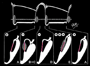
Mitral valve - papillary muscle complex during the
cardiac cycle.
|